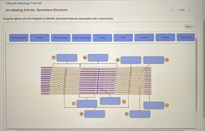
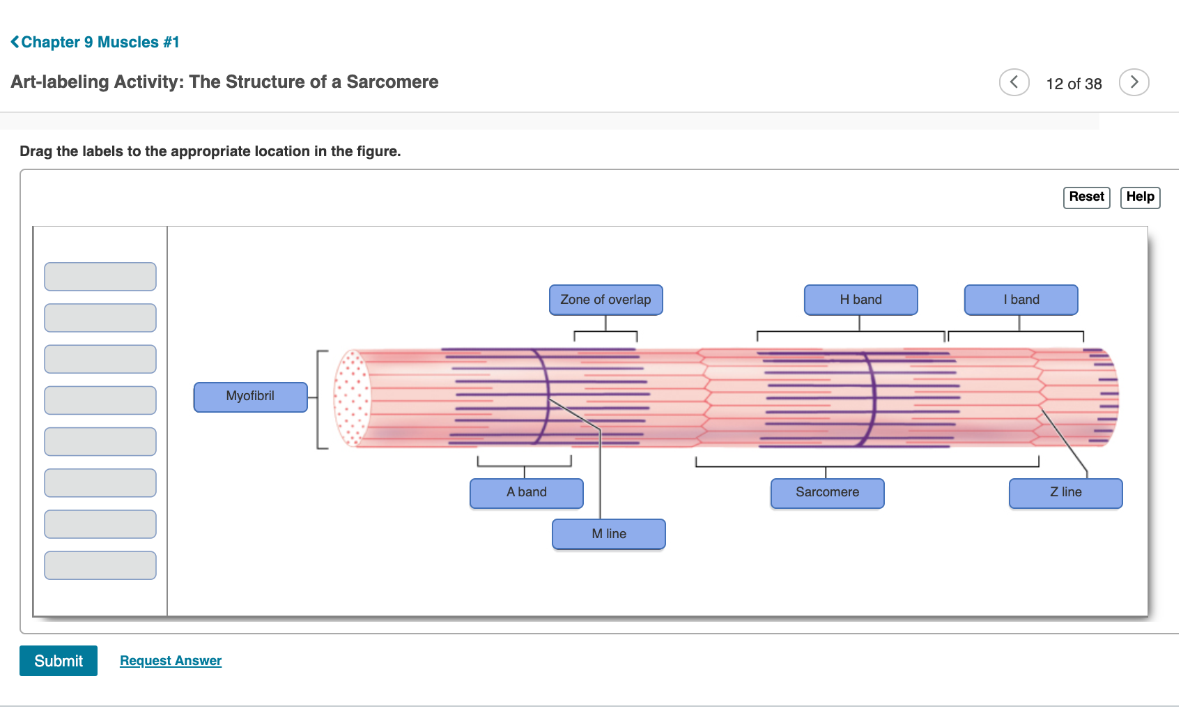
The Structure of a Sarcomere. Sarcomeres are connected to a plasma membrane called a sarcolemma by T-tubules which speed up the rate of depolarization within the sarcomere.

A P 1 Chapter 9 Mastering Assignments Flashcards Quizlet
Practice Test on Muscular Tissues.
. We describe cardiac muscle in detail in Chapter 18 but for easy comparison Table 93 on pp. For webquest or practice print a copy of this quiz at the Biology. Label different areas of an individual muscle unit known as a sarcomere below.
Reset Help Thick lament Thin filament Elastic ftamenti 1 band Mlino H zone Sarcomero Z disc Zone of overlap A band CCD WWW. Our focus here is to. A group of skeletal muscle fibers together with the surrounding perimysium form a n.
Multiple Choice Questions on Human Biology. Muscle Activity Answers answers is available in our digital library an online access to it is set as public so you can get it instantly. Each of the three muscle types has a structure and function uniquely suited to its function in the body.
Merely said the anatomy skeletal muscle activity answers is. A Lactic acid 7. ÎThe contractile unit of the myofibril is called the sarcomere.
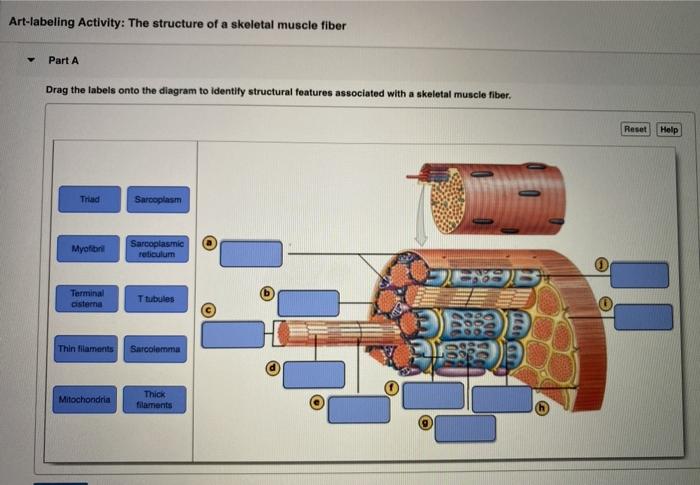
Structure of a Skeletal Muscle Fiber. The Structure of a Skeletal Muscle Fiber. A sarcomere is the basic unit of muscle tissue in both cardiac and skeletal muscle.
310311 summarizes the characteristics of all three muscle types. Î The epimysium penetrates and subdivides the muscle into muscle fiber bundles called the fascicles fasciculi. The structure of a skeletal muscle fiber.
Multiple Choice Questions on Animal Physiology. Then say My name is say a name that isnt yours and the muscle will test weak. The striated appearance of skeletal muscle fibers is due to the arrangement of the myofilaments of actin and myosin in sequential order from one end of the muscle fiber to the other.
D troponin tropomyosin and actin 5. Start studying Art-labeling Activity. In this chapter we first examine the structure and function of skeletal muscle.
D troponin tropomyosin and actin 5. Then we consider smooth muscle more briefly largely by comparing it with skeletal muscle. The muscle will test strong.
Learn vocabulary terms and more with flashcards games and other study tools. Which thin filament-associated protein binds two calcium ions. The Structure of a Sarcomere.
The storage and release of calcium ions is the key function of the. Learn vocabulary terms and more with flashcards games and other study tools. A sarcomere is defined as the region of a myofibril contained between two cytoskeletal structures called Z-discs also called Z-lines and the striated appearance of skeletal muscle fibers is due to the arrangement of the thick and.
The functional and complicated unit of striated muscles is. In this activity students will follow a procedure that instructs them to color and label 4 different sheets1 The neuromuscular junction2 The sarcoplasmic reticulum3 The sarcomere4 The cross-bridge cycleAs students color and label each they will also address each step of. B Ca and Mg ions 8.
Actin A Band Mline Z A. Sarcomeres appear under the microscope as striations with alternating dark and light bands. Structure of Skeletal Muscle Î Skeletal muscle is covered by a fascia called the epimysium.
Diagram and label a sarcomere including a thick filament thin filament A band H zone I band Z A. Muscles webquest print page. A Lactic acid 7.
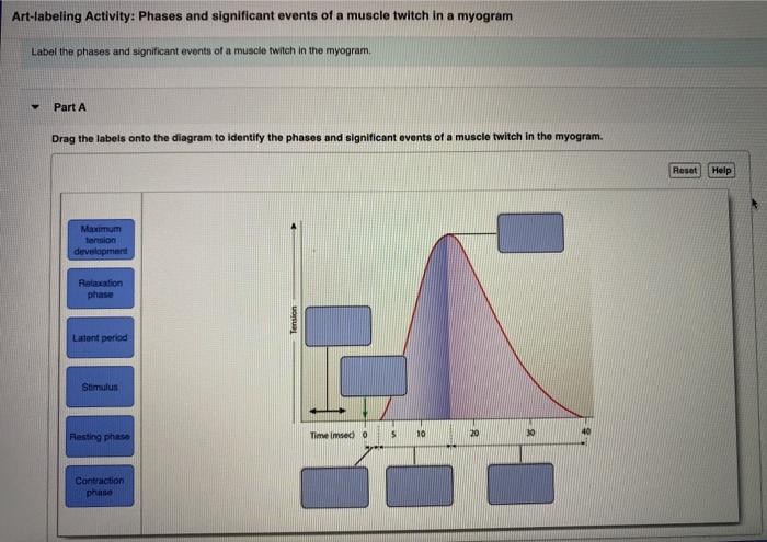
Î Each fascicle is covered by connective tissue called the perimysium. Chapter Test - Chapter 9 Question 3 Which thin-filament-associated structure is distinguished by its constituents of three globular subunits one of which has a receptor that binds two calcium ions. When the sarcomere contracts and shortens__________.
Multiple Choice Questions on Locomotion and Movement. Multiple Choice Questions on Muscle Contraction MCQ. The A band stays the same.
Start studying Art-labeling Activity. Each packet of these microfilaments and their regulatory proteins troponin and tropomyosin along with other proteins is called a sarcomere. Our books collection saves in multiple countries allowing you to get the most less latency time to download any of our books like this one.
The sarcomere is the contraction unit in the skeletal muscles that are under the control of motor. Read Free Muscle Test Questions And Answers muscle. Which of the.
The structure of a skeletal muscle fiber. Action potential propagation in a skeletal muscle fiber ceases when acetylcholine is removed from the synaptic cleft. Practice Test on Muscular System.
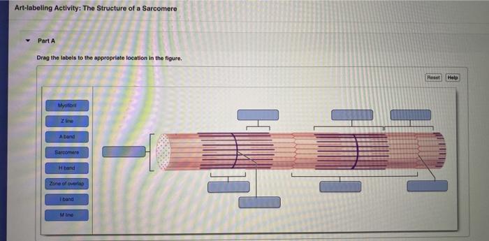
Instructors may assign this figure as an Art Labeling Activity using Mastering Figure 122 Relationship of the sarcoplasmic reticulum and T tubules to the myofibrils of skeletal muscle. The Microscopic Structure of a Myofibril H band Zone of overlap M line A band Sarcomere Z line I band Submit Previous Answers Request Answer Show more Biology Science Anatomy BIO 211. Structure and Bands of the Sarcomere Drag the appropriate labels to their respective targets.

Art Labeling Activity Sarcomere Structure Diagram Quizlet
Solved Muscle Histology Post Lab Art Labeling Activity Chegg Com

Art Labeling Activity The Structure Of A Skeletal Muscle Fiber Diagram Quizlet
Solved Art Labeling Activity The Structure Of A Sarcomere Chegg Com
Solved Art Labeling Activity The Structure Of A Sarcomere Chegg Com

A P 1 Chapter 9 Mastering Assignments Flashcards Quizlet
Solved Chapter 9 Muscles 1 Art Labeling Activity The Chegg Com
Solved Art Labeling Activity The Structure Of A Sarcomere Chegg Com
0 comments
Post a Comment